Image contest 2014 – The winners!
Light microscopy
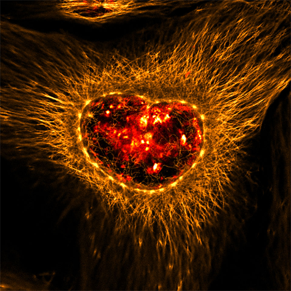 Ilse Oosterom (UMCU) ‘Burning the heart of pancreatic cancer’. The image shows chromosomal instability (CIN) in a pancreatic tumor cell derived from a genetically engineered mouse model for pancreatic cancer. The cell contains greater than two chromosomes as demonstrated by γ-tubulin staining (yellow). Dapi is shown in red and α-tubulin in orange. CIN in pancreatic cancer enables tumor cells to subvert restraints on unregulated growth.
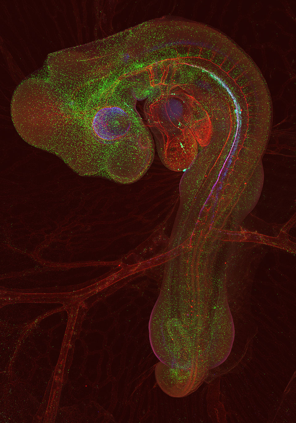
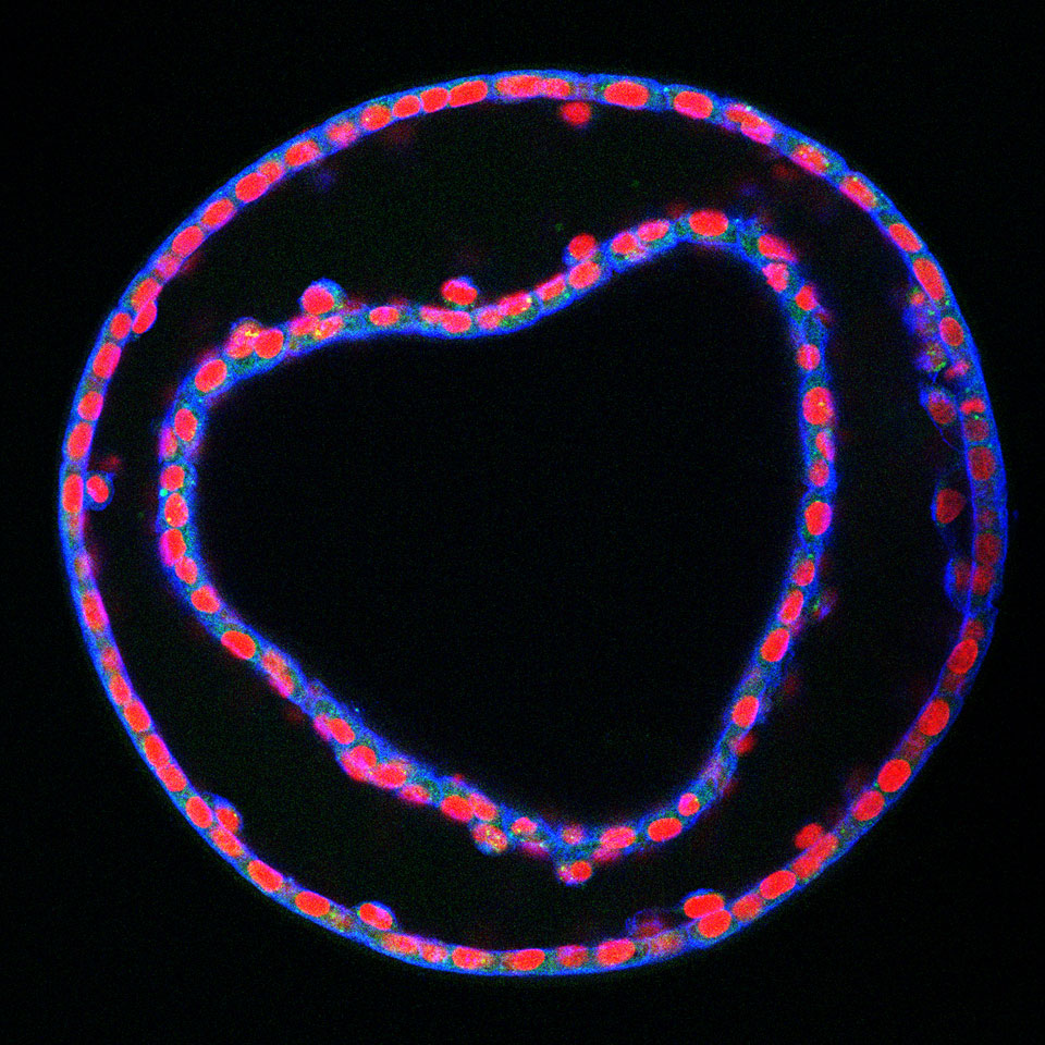 Marta Ruijter-Villani. “horse embryo heart”
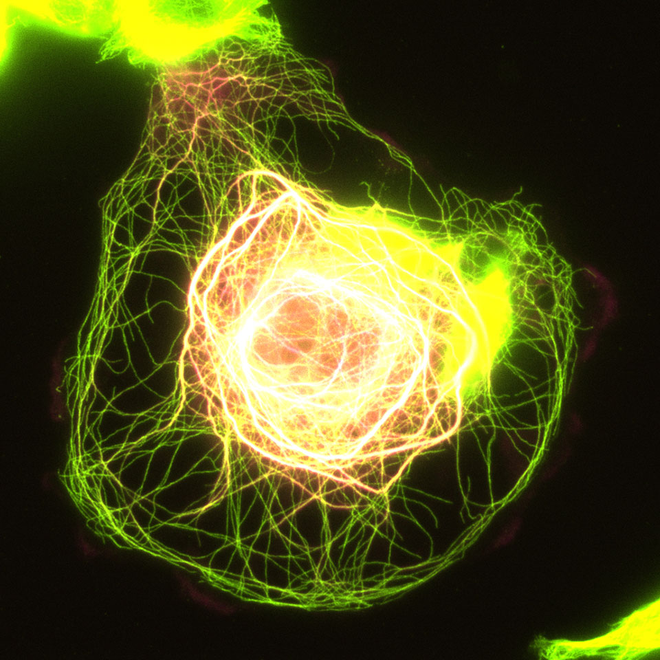 Ivar Noordstra (Cell Biology, UU). It is a breast cancer cell (MDA-MB-231) overexpressing CAMSAP2 (magenta). The tubulin staining (yellow) nicely shows the extreme effect of CAMSAP2 overexpression on the microtubule network. High levels of CAMSAP2 tend to bundle microtubules. In this particular cell the bundling was so strong that it looks like a giant electrical core with lightning reaching the membrane.
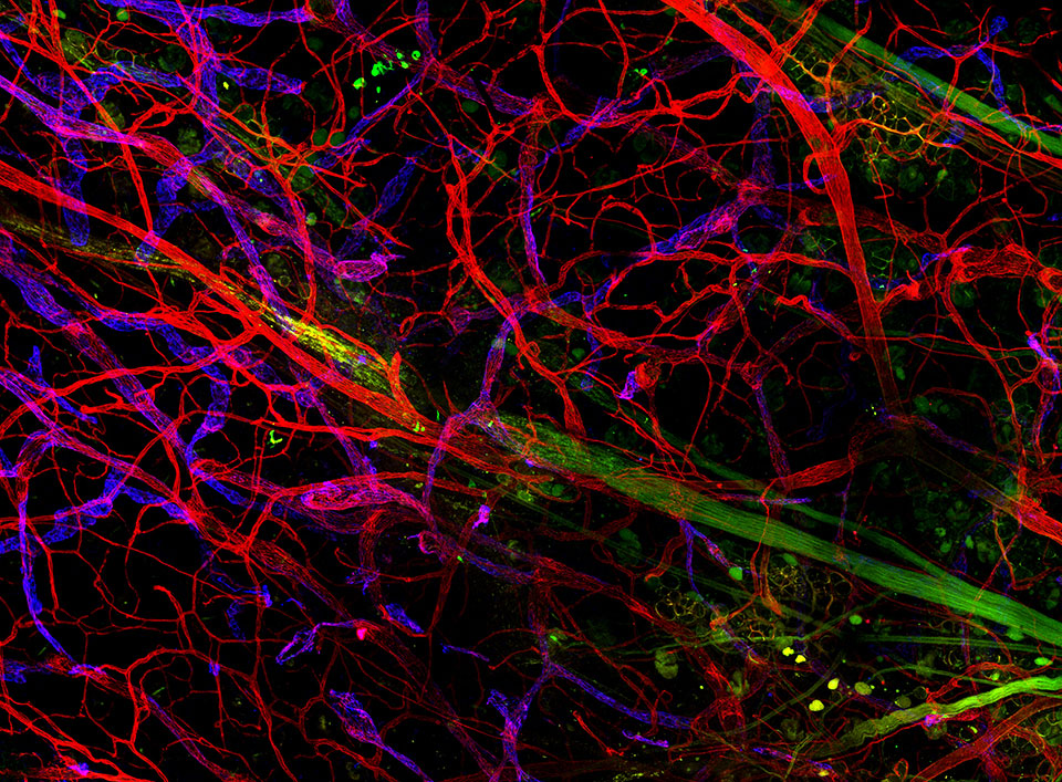 Serge van de Pavert (Hubrecht Institute). “Tangled up”. It is an image of a mouse ear stained for neurons, blood- and lymphatic- vessels.
 Dieudonnée van de Willige (Cell Biology, UU). It’s a picture of the axon terminus of a neuron transfected with a fill (in red) and a GFP-tagged protein that bundles actin and microtubules. It was made with the Nikon Eclipse 80i upright fluorescence microscope at the UU Cell Biology departement.
 Laurent Yvernogeau (Hubrecht Institute). “Three day-old chicken embryo surrounded by the yolk sac. The vessels (red) are stained with MEP antibody (secondary antibody coupled with Alexa-647), all hematopoietic cells (green) are stained with CD45 antibody (secondary antibody coupled with Alexa-555) and the hematopoietic precursors named hemogenic endothelial cells (blue) are stained with a Runx1 antibody (secondary antibody coupled with Alexa-488). The picture is a composite image of 70 panels, acquired on a LSM700 Zeiss confocal microscope (10x objective). Each z-step is 6.46μm (with a total of 162 z-stacks). The final composite picture was obtained using the Volocity software.”
Electron microscopy
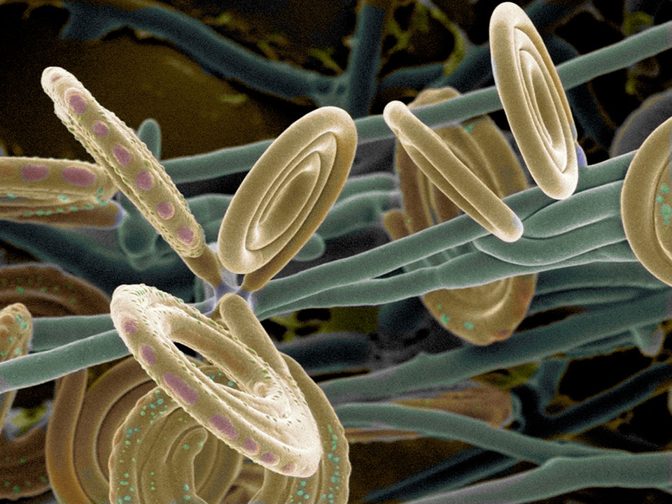 Jan Dijksterhuis (CBS Fungal Biodiversity Centre ) Helicosporon KLEIN
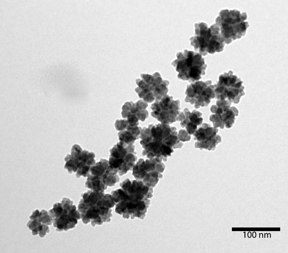
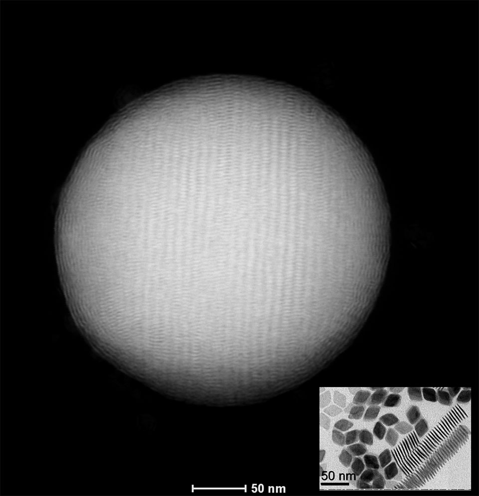 Da Wang (Debye Institute for Nanomaterials Science, UU) This is Da Wang from Soft Condensed Matter group. In my HAADF STEM image is the self-assembled “Supraball” composed of tens of thousands of rhombic shape GdF3 Nanocrystals, which is first time maded by our group. You can clearly see the orientation(s) and packing of the nanocrystals. The inset is the TEM image of the rhombic shape GdF3 Nanocrystals. Instrument: Tecnai 20F (FEI company), in HAADF-STEM mode Image title: The Beauty of Symmetry Content: Self-assembled “Supraball” composed of tens of thousands of rhombic shape GdF3 Nanocrystals Highlight: Highly ordered structure and symmetric. The supraball is first time found by our group.
 Akshatha Mohan. The image shown is of cauliflower shaped nanoparticles, taken with the Technai 10 microscope. The silicon nanoparticles are made in a single step in the gas phase by plasma enhanced chemical vapor deposition (PECVD). HRTEM imaging further shows that these are made of “smaller subsystems” of 2-5 nm. Silicon being abundant and non-toxic, makes silicon nanoparticles a good choice of materials for biodegradable in-vivo applications to diagnose and treat diseases. Silicon nanoparticles also are highly potential candidates for bio-markers in tissue imaging. In addition to their biological applications, silicon nanoparticles have a wide range of optical and light emitting applications enabling their utilization in lithium ion batteries, catalyzers in water splitting application and solar cells.
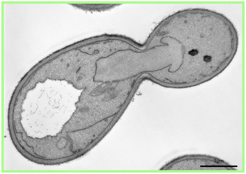 Despina Xanthakis (Cell Biology, UMCU) “One Direction” Het is een delende gist cel en de kern vormt een pijl welke naar de nieuw geboren cel wijst.
Doing Microscopy?
Have an image?
Win the prize!
For the third year the Bio-Imaging Utrecht workgroup organizes a microscope image competition. The focus of this competition is to show that microscopic images often not only have scientific value but also an artistic value, the images will be judged on their visual effect.
Any microscope image can participate in the competition. We have two categories: Fluorescence images and Electron microscopy images. The first price will be a 200,-gift certificate sponsored by Carl Zeiss BV.![]()
A few rules apply:
- One image per person can be submitted.
- Mild processing and photo shopping is allowed.
- The image must be made with equipment present on the Uithof.
Image format: any!
Image size: any!
Deadline: October 15th, 2014
How to participate: send your image (with short description) to meetings@bioimaging-utrecht.nl
Utrecht Microscopy Image Contest 2014
Livio Kleij (Molecular Cancer Research, UMCU)
Anko de Graaff (Hubrecht imaging centre, Hubrecht)
Ilya Grigoriev( Cell Biology, UU)
Corlinda ten Brink (Cell Biology, UMCU)
Dave van den Heuvel (Molecular Biophysics, UU)