Image contest 2016 – The winners!
Light microscopy
 Anne Gelderloos / University Utrecht
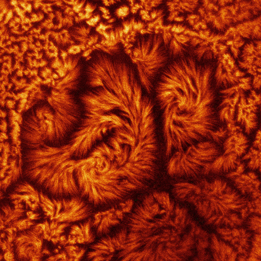
The artistic name of the image: Burning Brush Border
Description: Microvilli of the Caco-2 brush border visualized by actin staining.
Microscope: Leica TCS SP8 STED
 Youri Adolfs / University Utrecht Medical Center
Artistic title: The rewarding and sympathetic development
Scientific description: This is a E12.5 whole mouse embryo which was stained for Tyrosine hydroxylase (TH). TH which catalyzing the conversion of L-tyrosine to L-DOPA is depicted here in green. In order to image the embryo as a whole, the embryo was made transparent, with the clearing method 3Disco. To show the whole structure of the embryo, as a kind of counterstaining, an image of the autofluorescence of the embryo (depicted in blue) was made. The TH staining clearly shows how the axon bundles of dopaminergic neurons within the midbrain, runs from the midbrain towards the forebrain (medial forebrain bundle). Outside of the CNS, TH stains the developing sympathetic nervous system.
Microscope: Light Sheet microscope: UltraMicroscope II (LaVision BioTec)
 Michele Fedecostante / University Utrecht
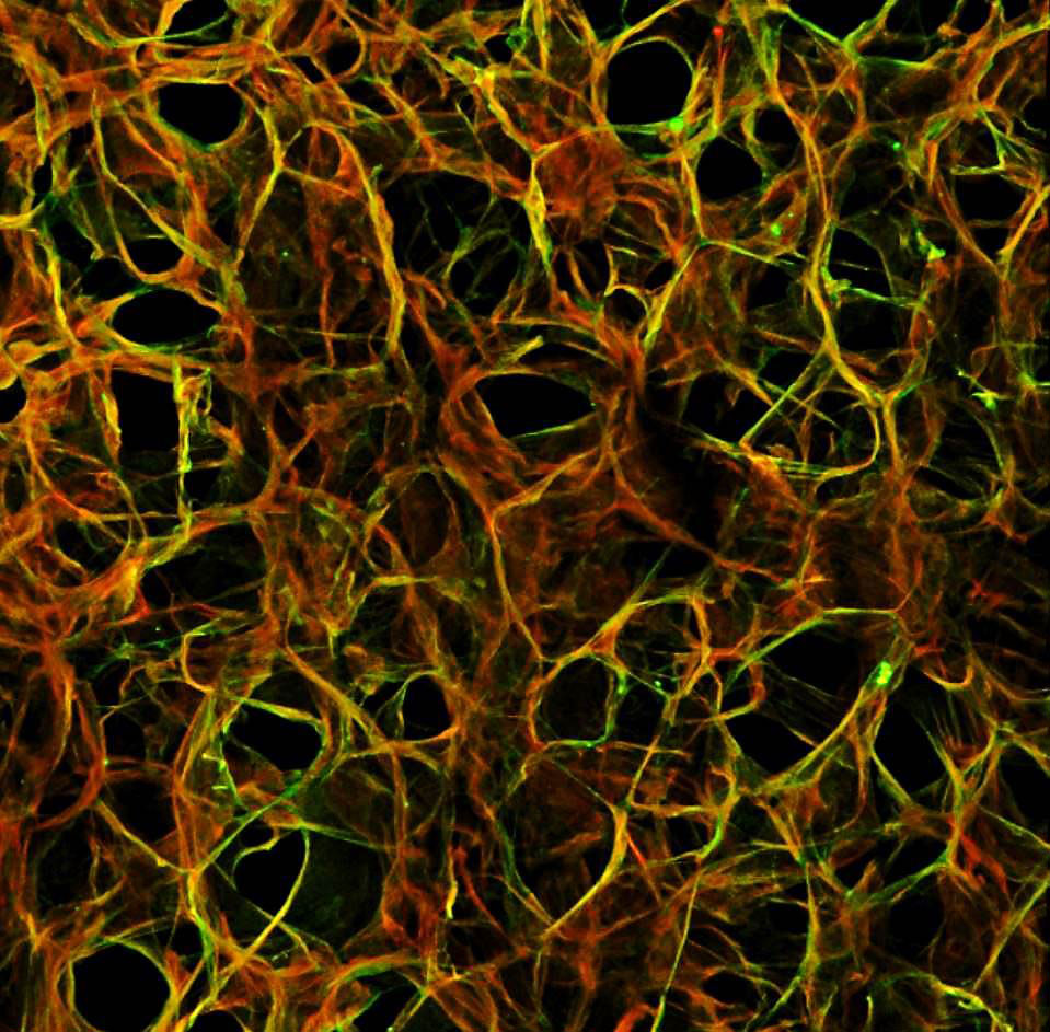
Artistic title: Science is all about network
Scientific description: A whole rat kidney has been decellularized, using an SDS-based protocol to obtain a cell-free kidney scaffold. 150um cryosections have been obtained and processed for immunofluorecence. collagen IV (green) and laminin (red) are the main proteins characterizing the extracellular matrix. The tight network shows that the scaffold preserved a proper structure after the decellularization protocol.
Microscope: Leica SPE-II confocal microscope
Electron microscopy
 Jan Dijksterhuis / CBS-KNAW Fungal Biodiversity Centre
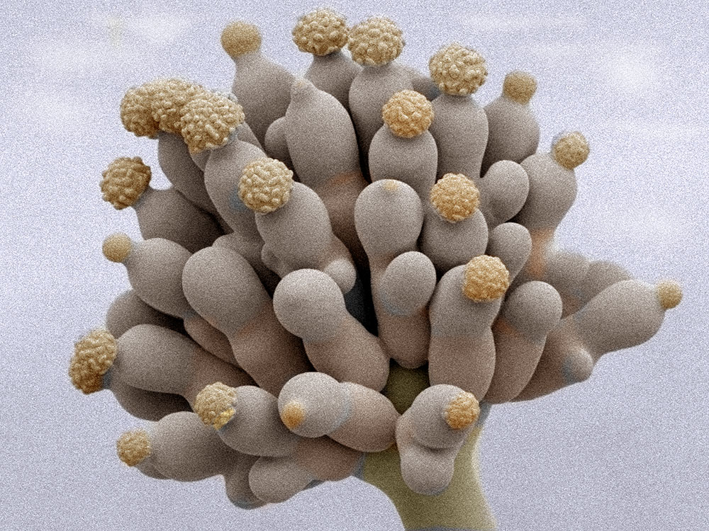
Artistic title: Aspergillus mutant, making spores in a different way
Microscope: JEOL 5600 LV SEM equipped with an Oxford CT1500 Cryostation.
Scientific description: Aspergillus mutant, making spores in a different way. It appears as a flower from another planet, useful for science fictions movies to come. The ornamented spores have a size of approximately 4 µm. The view on the spore-forming structures is unusual clear here.
 Job Fermie / University Utrecht Medical Center
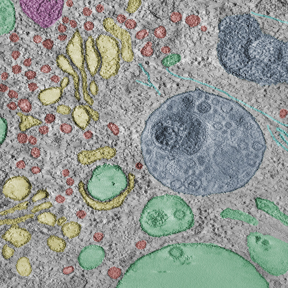
Artistic title: Exploring the cellular landscape
Microscope: FEI Tecnai T20 in the CMC and processed in IMOD
Scientific description: Hela cells were high-pressure frozen and embedded in Lowicryl HM20 resin. A tomogram was generated from a 300 nm thick section of these embedded cells. A variety of organelles and structures is seen, including early and late endosomes (green and blue respectively), microtubules (cyan) and golgi stacks (yellow) with their associated vesicles(red).
 Sergey Loginov / University Utrecht
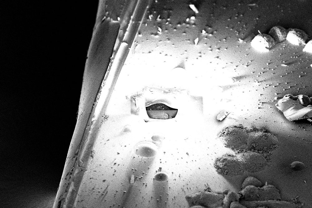
Artistic title: Aftermath
Microscope: FEI Scios
Scientific description: This is a SEM image of a sample after the 3D data acquisition by the FIB was finished. Sample: SKBR3 cells.