Image contest 2018 – The winners!
Light microscopy
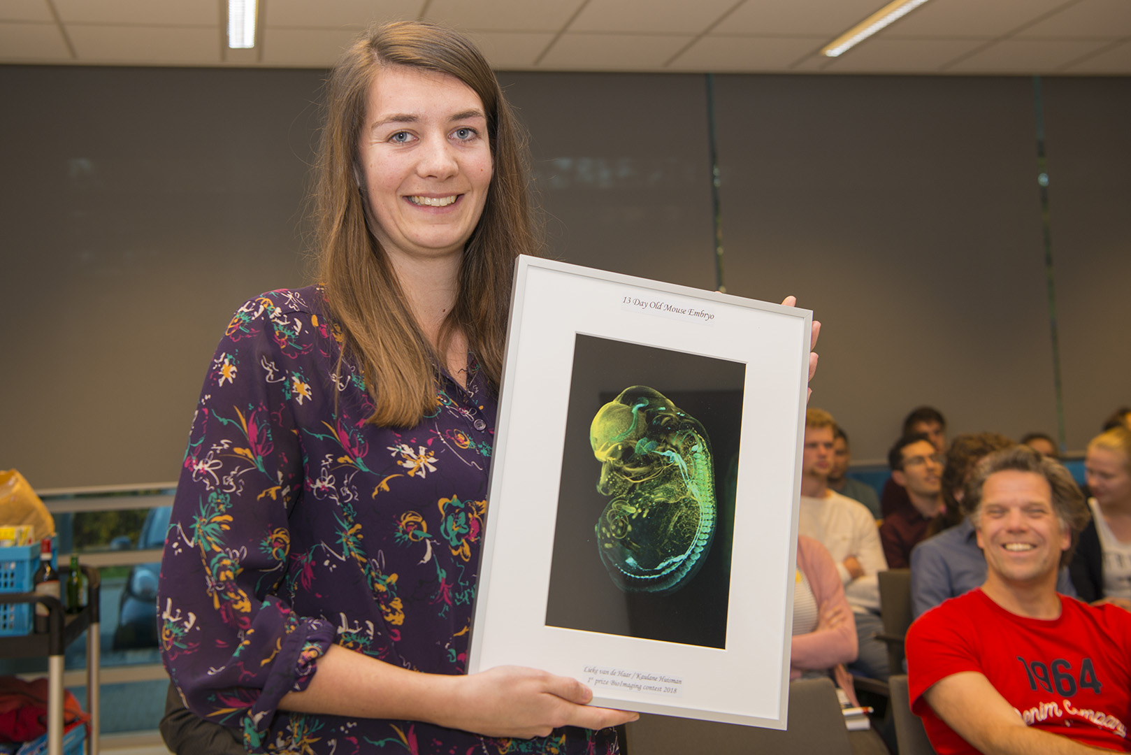
 Lieke van de Haar / Kaulane Huisman, UMC
The artistic name of the image: 13 day old mouse embryo
Description: 13 day old mouse embryo showing the development of the dopaminergic system and Neuropilin 2 positive neurons in the olfactory bulb and the habenula. The embryo is prepaired with 3DISCO staining and imaged using a LaVision Light Sheet UltraMicroscope.
Microscope: : LaVision Light Sheet UltraMicroscope
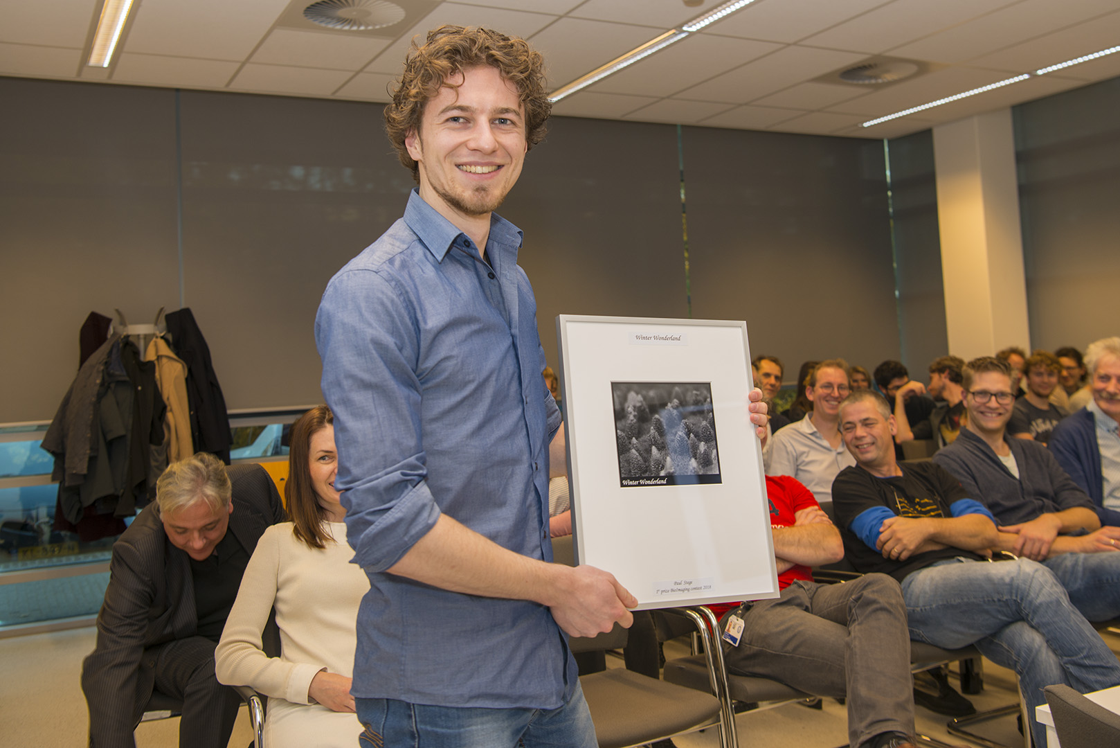
The winners award ceremony!:
 Jasper & Eitan Zahavi, UU Cell Biology
Artistic title: Tryptich
Description: Hippocampal neurons grown in micropatterened devices to compartmentalize distal axons. GFP (Green), Tau (Red), DNA (Blue).
Microscope: Confocal Zeiss LSM700
 Marijn Peters, UMC
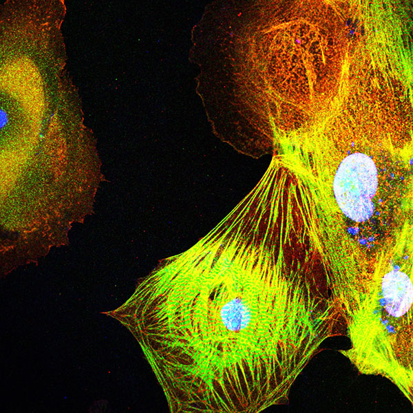
Artistic title: Structural changes in ischaemic human iPS cardiomyocytes
Scientific description: 63x magnification image of human induced pluripotent stem cell derived cardiomyocytes (iPS-CM) after 24 hour incubation in a low oxygen concentration. This image was made after double staining for cardiomyocyte sarcomeric proteins alpha-actinin (green) and troponin T (red) to identify the cardiomyocytes that maintain sarcomere integrity and the cardiomyocytes that lose sarcomere organization even though the alpha actinin and troponin T proteins are still expressed. DAPI was used as a nuclear marker.
Microscope: Confocal Leica SP8x
Electron microscopy
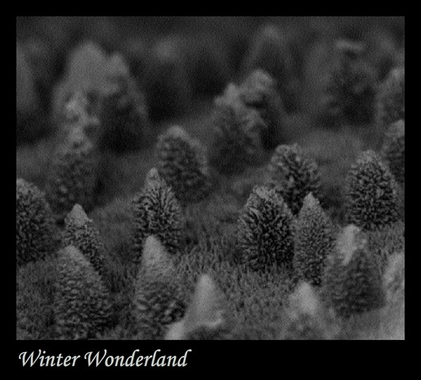 Paul Stege, UMC
Artistic title: Winter Wonderland
Microscope: Phenom Desktop SEM
Scientific description: A 7000x magnification of the lambrum of a damselfly.
The winners award ceremony!:
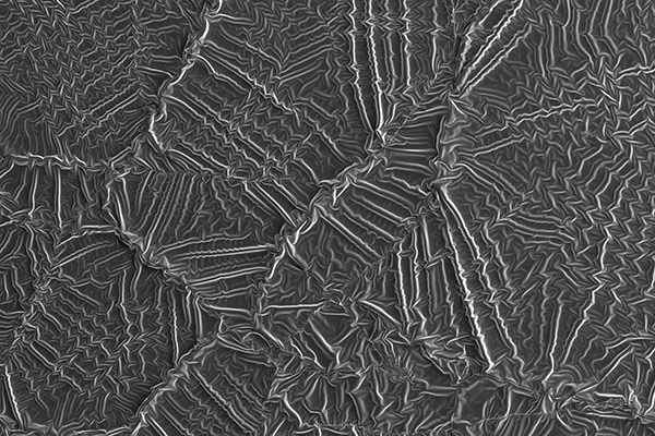 Maarten Bransen, UU
Artistic title: Fingerprint of a polymerizing droplet.
Microscope: Fei Helios DualBeam g3-uc
Scientific description: the image (ca. 800×500 μm) shows the surface of a droplet of a dense suspension of gold nanoparticles after solidifying by means of a polymerization reaction of the solvent. When the polymerization reaction is initiated with a strong UV pulse, it can lock the particles in place, thus preserving their distribution in the liquid. Sometimes, a thin dried-out layer is present at the surface of the droplet when the reaction is initiated. As the material contracts and shrinks during the reaction, this surface layer wrinkles and folds, giving rise to beautiful wave-like patterns over many length scales.
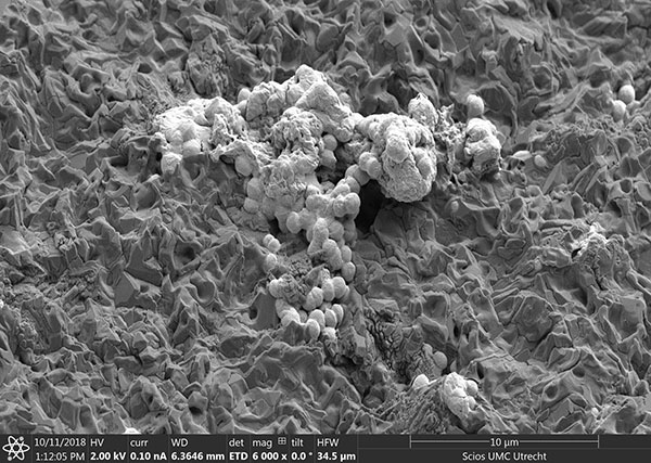 Marco Viveen, UMC
Artistic title: Snow in the Desert
Microscope: FEI Scios
Scientific description: Enterococcus faecium grown on blood agar plate. Fixed with and dehydrated for imaging with the FEI Scios (from the cell biology).