2016 Seasonal Utrecht Microscopy Meeting
Fall Meeting: Tuesday November 29th
- UMC – Stratenum Building – Vondelzaal (Rm 3.108) 15.00 – 17.00, drinks afterwards
- “Lies, damn Lies, and Image Analysis”, Gerhard Blab, Utrecht University
- “Come and compare the small microscopes out there”
– Cytell imaging system from GE Healthcare presented by Chromaphor
– DMI1 microscope from Leica
– Evos imaging system from Thermo Scientific
– IX83 microscope from Olympus
– Zoe imaging system from Biorad(Demo only)
– These systems will be available for demo and hands on during the entire day - BioImagingUtrecht Image contest: The winner of the 2016 BioImaging Utrecht Image contest will be announced at the end of the meeting.
- Program, click here (pdf).
Additional activities
- ImageJ beginners workshop: October 11th
- Image competition 2016 (send images in October)
- Special seminars that will be announced separately
- 2016 overview, click for pdf.
UU launches new STED microscopy facility
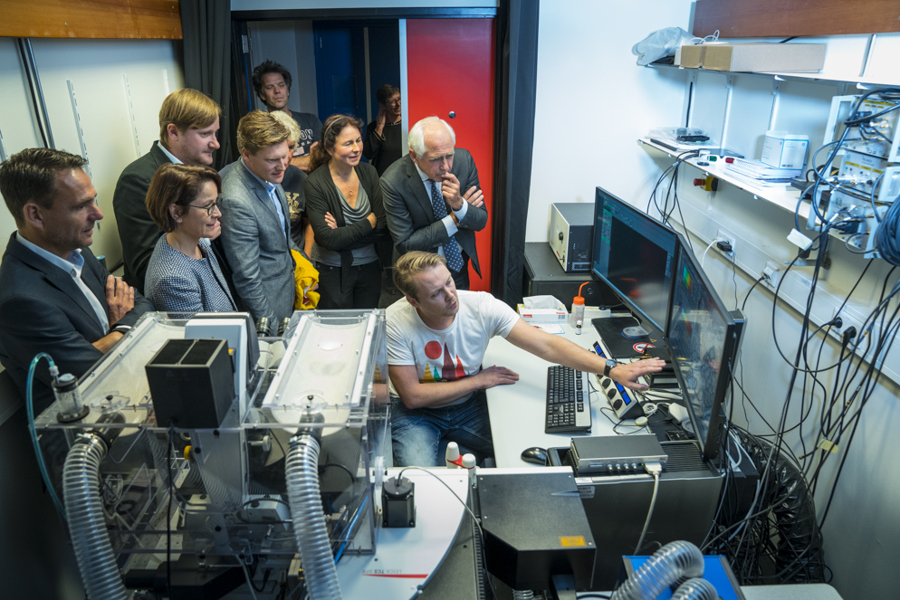 On 27 October Bert van der Zwaan, Rector of Utrecht University and Eva van Pelt, Vice President Sales EMEA , Leica, officially opened the STED (STimulated Emission Depletion) microscopy facility at the Biology Imaging Center of the Department of Biology. STED microscopy facility will enable researchers working in the area of Life Sciences to study the architecture and dynamics of cellular components at a resolution beyond the diffraction limit.
Utrecht University offers a unique research infrastructure, which is amongst the best of its kind in Europe. It plays a significance role for academic research. The Biology Imaging Center provides access, support and training in advanced light and fluorescent microscopy techniques for research groups within the department of Biology and also to groups from other institutes within and outside Utrecht.
The STED microscopy facility, which is part of the Biology Imaging Center, is a nice example of collaboration in the development of instruments and their applications. Collaborating partner of the STED microscopy facility is Leica.
Opening of STED microscopy facility at Utrecht University
October 27, Utrecht University (Kruytbuilding room O622, Padualaan 8, Utrecht; pdf)
- Scientific Session
14:00-14:30 Lukas Kapitein, Cell Biology, UU “Unraveling cytoskeletal organization using optical nanoscopy”
14:30-15:00 Hans Gerritsen, Molecular Biophysics, UU, “Fluorescence Lifetime Imaging using STED”
15:00-15:30 Nathalie Garin, Leica, “STED microscopy: new horizons for life scientists”
15:30-16:00 Fedja Bobanovic, Leica, “Leica STED: From Technology to Applications”
16.00-16.15 Coffee break
- Official Opening
16.15-16:20 Welcome by Anna Akmanova, Professor of Cell Biology, UU
16.20-16.40 Eva van Pelt, Vice President Sales EMEA , Leica, “The role of public-private collaboration in the development of instruments and their applications ”
16.40-17.00 Bert van der Zwaan, Rector of Utrecht University “The significance of infrastructures for academic research”
17.00-17.15 Tour of the facility
17.15 Drinks
Image analysis for beginners using ImageJ (free multiplatform software): Tuesday October 11th (BBG175, BuysBallot Building)
- If you want to start using ImageJ/FIJI and have no clue where to begin. Or you just started using it and got stuck.
- During the hands-on session our experts will guide you through your specific problem.
- For the hands-on: please fill in the registration form and send it to meetings@bioimaging-utrecht.nl. (The talk can be attended without registration.)
- Deadline for registration is the 1st of October. Be quick: the number of spots for the hands-on is limited.
- Program (pdf):
12:15 -13.00 Talk: Introduction to the basics of image analysis
13.00 -15:00 Hands-on session 1
15:00 -15:15 Coffee break
15:15 -17:00 Hands-on session 2
Spring meeting: May 24th (Stratenum)
Intravital Imaging: Microscopy and spectroscopy in live animals
- “Insights into cell division using C. elegans”, Matilde Galli, Hubrecht Institute
- “The zebrafish embryo as in vivo developmental and functional tool”, Federico Tessadori, Hubrecht Institute
- “Use of intravital imaging to understand the dynamic nature of cancer”, Maria Alieva Krasheninnikova, Hubrecht Institute
- Program, click here (pdf).
Winter Meeting Tuesday February 2nd
- UMC – Stratenum Building – Vondelzaal (Rm 3.108) 15.00 – 17.00, drinks afterwards
- Bas Cloin (Utrecht University) “Adaptive optics for super-resolution microscopy”
- Marvin Tannenbaum (Hubrecht Institute) “Translational dynamics on single mRNA molecules in vivo”
- Program details, click here (pdf).